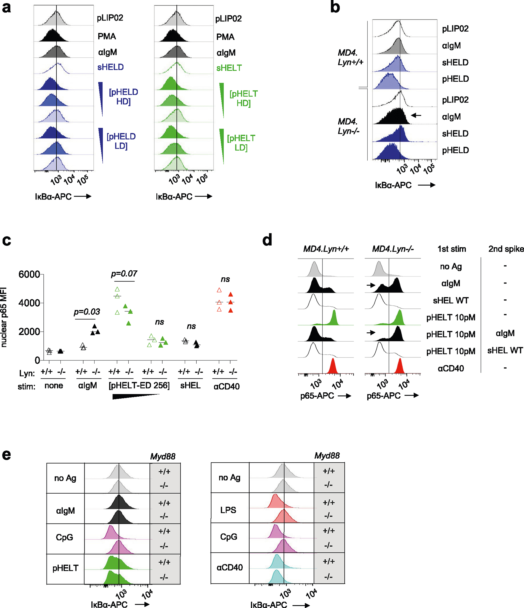
Extended Data Fig. 6 |. pHEL robustly trigger NF-κB independently of MYD88.

a. As in Main Fig. 7b-d except histograms showing IκBα degradation at 20 minutes post-stimulation for soluble stimuli sHELD/T and pHELT/D with high or low ED (HD vs. LD). Inset triangles represent decreasing concentration of pHEL (10, 1, 0.1 pM). Data are representative of >= 4 independent experiments. b. As in Main Fig. 6b, except splenocytes from Lyn+/+ and Lyn–/– MD4 mice were stained to detect intracellular IκB in CD23+ B cells at 20 minutes. Data are representative of 3 independent experiments. c. Graph corresponds to data in Main Fig. 7g,h except nuclear p65 MFI rather than % p65 positive nuclei are quantified. Graph depicts data with mean from 3 independent experiments. Groups were compared by paired two-tailed parametric T-tests. d. Nuclear p65 translocation is detected as in Fig. 7f except stimuli are applied on ice for 15 min each, first 10pM pHELT-HD +/− superimposed soluble stimuli (anti-IgM 10 μg/ml or sHEL-WT 1 μg/ml), followed by 20 min 37 C incubation as in Main Fig. 6f. e. As in Main Fig. 4c,d except stained to detect intracellular IκB in CD23 + B cells at 20 minutes. Data are representative of 3 independent experiments. In panels A, B and E, line in offset histograms references internal negative control.
