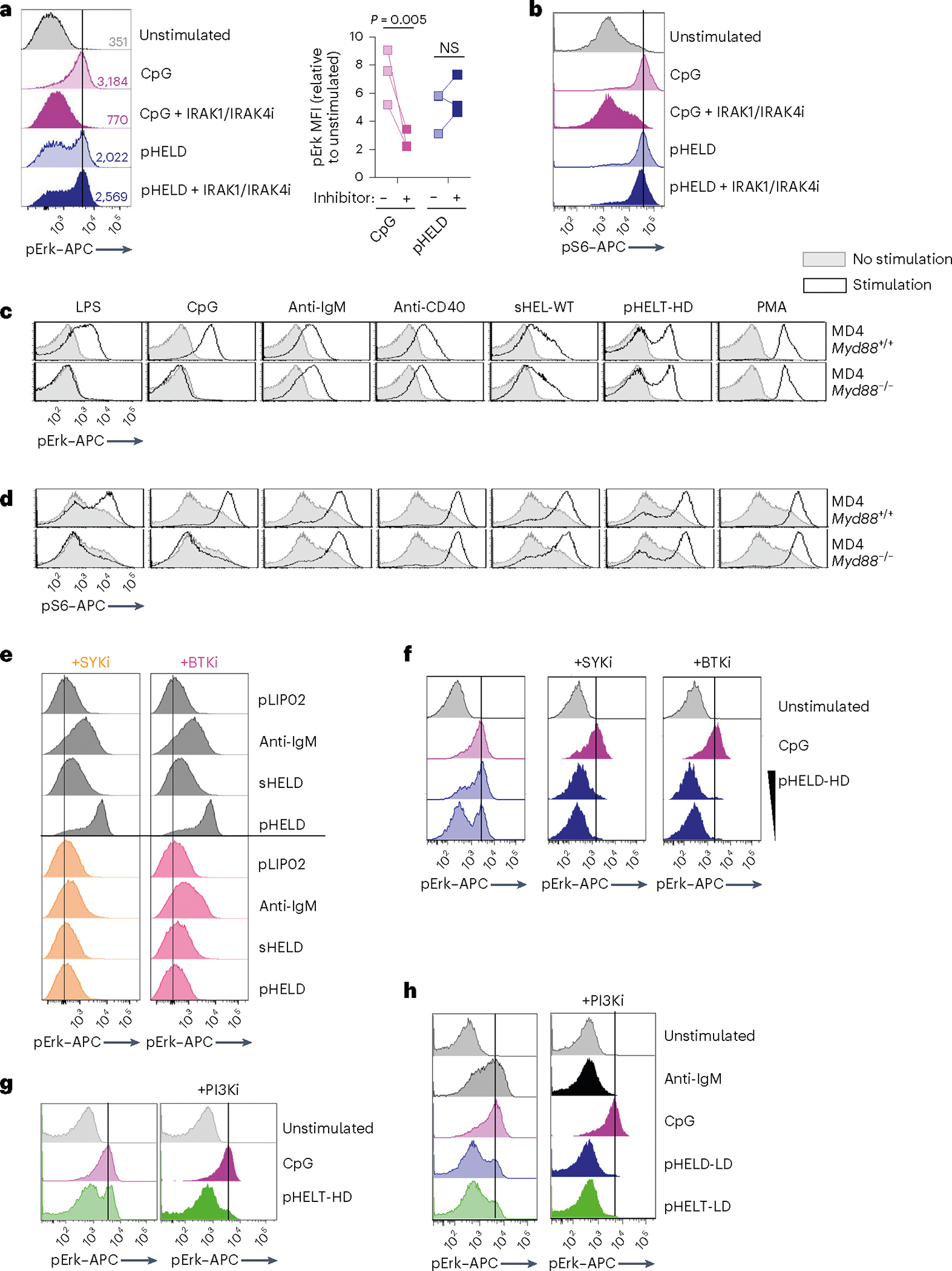
Fig. 4 |. pHEL signaling is independent of MYD88/IRAK1/IRAK4 but requires the canonical BCR pathway.

a,b, Pooled splenocytes and lymphocytes from MD4 mice were preincubated at 37 °C for 15 min with Rigel R568 IRAK1/IRAK4 inhibitor (IRAK1/IRAK4i; 1 μM), subsequently incubated for 20 min with the indicated stimuli (2.5 μM CpG and 1 pM pHELD), and then fixed, permeabilized and stained to detect pErk or pS6 as well as B220. Representative histograms depict pErk (a) or pS6 (b) in B220+ B cells. The graph depicts pErk MFI from three independent experiments. Data in b are representative of at least four independent experiments. Data were compared by unpaired two-tailed parametric t-test. c,d, Pooled splenocytes and lymphocytes from MD4 Myd88+/+ and MD4 Myd88−/− mice were stimulated with the indicated stimuli for 20 min and stained to detect pErk or pS6 in B220+ B cells as in a and b (10 μg ml−1 lipopolysaccharide (LPS), 2.5 μM CpG, 10 μg ml−1 anti-IgM, 1 μg ml−1 anti-CD40, 1 μg ml−1 sHEL-WT or 1 pM pHELT-HD (ED of 256)). Histograms depict pErk (c) or pS6 (d) and are representative of four (c) or three (d) independent experiments. e,f, As in a except with pretreatment with SYK inhibitor (SYKi; 1 μM Bay 61–3606) or BTK inhibitor (BTKi; 100 nM ibrutinib), followed by a 20-min incubation with the indicated stimuli: 1 pM and 0.1 pM (f) pHELD-HD (ED of 286), 1 μg ml−1 sHELD, 10 μg ml−1 anti-IgM and 2.5 μM CpG. Histograms are representative of four independent experiments. g,h, As in a except pretreatment with PI3K inhibitor (PI3Ki; 10 μM Ly290049) and stimuli (1 pM pHELT-HD ED = 256 in g; 1 pM pHELD-LD ED = 53 and pHELT-LD ED = 58 in h). Histograms are representative of four independent experiments.
