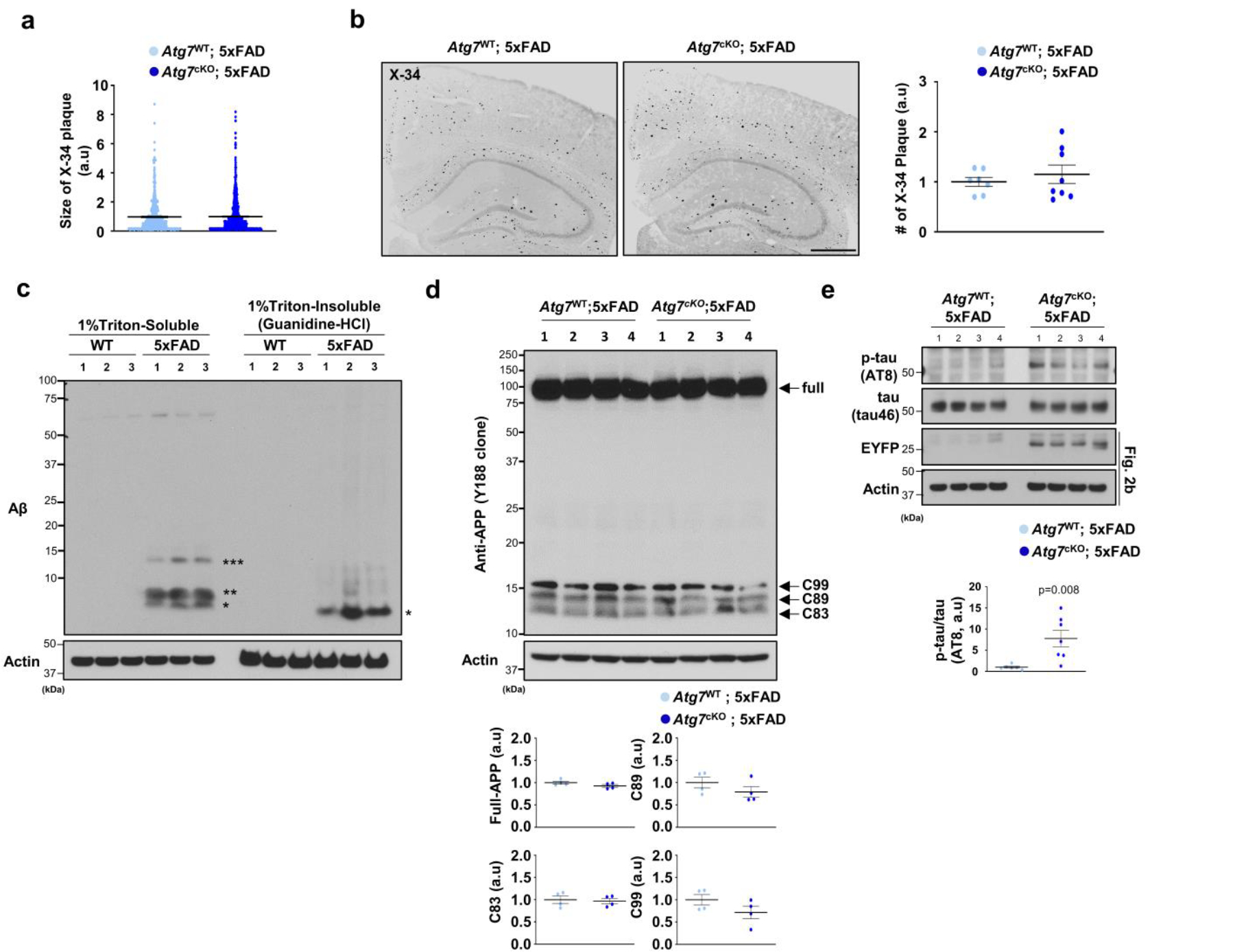
Extended Data Fig. 3 |. Analysis of levels of full-length APP, C-terminal fragments (CTF), and Aβ, tau, and the number and size of amyloid plaques.

(a) The size of X-34+ amyloid plaques was quantified from 489 plaques for Atg7cKO; 5xFAD (7 male mice) and 763 plaques for Atg7WT; 5xFAD (4 male mice). Scale bar, 10 μm. (b) The number of X-34+ amyloid plaques was quantified from Atg7cKO; 5xFAD (8 male mice) and Atg7WT; 5xFAD (7 male mice). Scale bar, 500 μm. (c, d) Brains from 5xFAD mice (3 female, c) and littermate controls (3 female) or Atg7cKO; 5xFAD mice (4 male mice, d) and Atg7WT; 5xFAD mice (4 male mice) were homogenized, fractionated into 1% Triton x-100 soluble and insoluble fractions, and processed for Western blot using antibodies against amyloid-beta (c) and APP (Y188 clone) (d). (e) Brains from Atg7cKO; 5xFAD (7 male mice) and Atg7WT; 5xFAD mice (6 male mice) were homogenized in 1% Triton x-100. Levels of amyloid-beta, p-tau (AT8), and tau were examined through Western blot. EYFP indicates the presence of Cx3crCreER in the mice that co-express EYFP and CreER. Actin was used as a loading control. p = 0.008. p-values were calculated by unpaired two-tailed Student’s t-test. All values are reported as mean ± SEM. Source numerical data and unprocessed blots are available in source data.
