. 2024 Mar 19;12:RP88866. doi: 10.7554/eLife.88866

Figure 1. Discovery of apicoplast membrane proteins.

Activity test of biotinylation of proximal proteins by APT1-TurboID-4Ty. Parasites were grown in 500 μM D-biotin for 90 min, followed by detection of biotinylated proteins (green) by streptavidin reagents on indirect fluorescence assay (IFA) (A) and on western blots (B). Actin served as the loading control. Scale = 5 μm. (C) Volcano plot analysis comparing the TurboID fusion to the parental line. Three replicate mass-spectrometry experiments were analyzed by the Student t-test. Known apicoplast proteins (red) and candidates with different numbers of transmembrane domains (TMDs; deep and light blue were indicated, and novel apicoplast proteins were pointed out; see Supplementary file 2a). (D) Workflow of the discovery of novel apicoplast membrane proteins. Candidates from hyperLOPIT (Barylyuk et al., 2020), ACP-BirA (Boucher et al., 2018), and APT1-TurboID (this study) were screened by epitopte tagging and IFA. (E) Confocal co-localization of HAP-6Ty with the apicoplast marker ACP, showing confocal imaging of HAP-6Ty (red) and ACP (green), and Pearson correlation coefficiency (PCC). The PCC values over the merged fluoresent foci for candidate proteins and ACP were analyzed by a co-localization and region of interest (ROI) intensity analysis module in the NIS Elements AR software and shown with mean ± standard error of the mean (SEM; N = 6 for each). Scale = 5 μm. (F–H) Summary and comparison of the screening from three datasets. The putative transporters are grouped into diverse types (F) and identified from three different datasets (G), which shared three novel proteins, as shown by the Venn diagram (H). Three independent experiments were performed with similar outcomes (A, B, E).

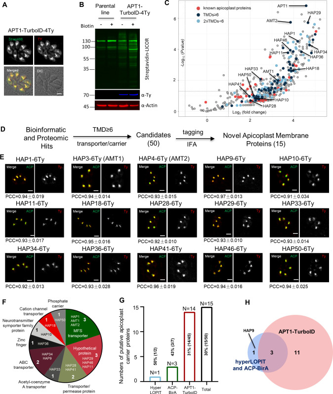
Figure 1—source data 1. PDF file containing the uncropped western blot gels for verification of the TurboID fusion lines in Figure 1A.
Figure 1—source data 2. Excel file containing the raw images of confocal analysis for verification of co-localization of newly discovered proteins with the apicoplast marker ACP.

Figure 1.

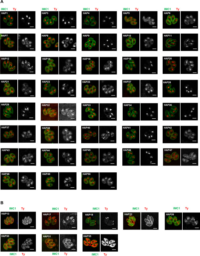
Figure 1—figure supplement 1. Indirect fluorescence assay (IFA) analysis of HAP-6Ty fusions in parasites.

Figure 1—figure supplement 1.

(A, B) Candidates from hyperLOPIT, ACP-BirA, and APT1-TurboID were endogenously tagged with 6Ty at their C-termini using a CRISPR-Cas9 approach, followed by IFA analysis with antibodies against the inner membrane complex protein IMC1 (green) and Ty (red). Many of the HAPs were localized to a foci similar to position of the apicoplast in parasites (A), whereas others were localized to the cytosolic vesicles (e.g. HAP30, HAP13, and AP35), pellicular membrane (e.g. HAP22), or other structures (B). Scale = 5 μm. Note that HAP2, 15, 24, and 39 were not successfully tagged for IFA analysis. Three independent experiments were performed with similar outcomes and representative images are shown.
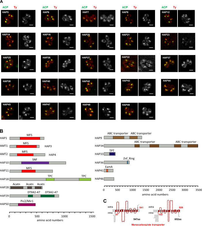
Figure 1—figure supplement 2. Analyses of non-apicoplast-localized HAP fusions, and domain analysis of novel apicoplast transporters.

Figure 1—figure supplement 2.

(A) Shown here are the HAP-Ty fusions that were not localized to the apicoplast. Indirect fluorescence assay (IFA) analyses were performed with the HAP-6Ty fusions (red) and the apicoplast marker ACP (green). Scale = 5 μm. (B) Domain analysis of newly identified apicoplast membrane proteins. Proteins were analyzed by IntroProScan (https://www.ebi.ac.uk/interpro/about/interproscan/). MFS, major facillitator transporter superfamily capable of transporting small solutes in response to chemiosmotic ion gradients (Pao et al., 1998; Walmsley et al., 1998); SNF, sodium:neutrotransmitter symporter superfamily that is responsible for exitatory amino acid transport (Malandro and Kilberg, 1996); TPC, two-pore channels that have roles in organelle integrity, inter-organelle communication and growth in T. gondii (Li et al., 2021); acatn, acetyl-coenzyme A transporter (Bora et al., 1999); DTX42-47, among which DTX43 is a citrate transporter responsible for loading citrate into xylem tissues and facilitate iron transport to shoots (Green and Rogers, 2004; Rogers and Guerinot, 2002); ABC, ABC transporters that are involved in export or import of a wide variety of substrates ranging from small ions to macromolecules; Pic2/Mir1-like, phosphate carriers that were reported to import copper and inorganic phosphate into mitochondria (Hamel et al., 2004; Vest et al., 2013); TPT, Sugar phosphate transporter domain with a specificity for triose phosphate (Jack et al., 2001); Znf ring, Zinc finger that may bind metals, such as iron, or no metal at all; EamA, found in many members classed as drug/metabolite transporters (Jack et al., 2001). Protein lengths were scaled by amino acid numbers (aa). (C) The topology of two monocarboxylate transporters (AMT1 and AMT2) was analyzed by an online server PROTTER (https://wlab.ethz.ch/protter/start/).