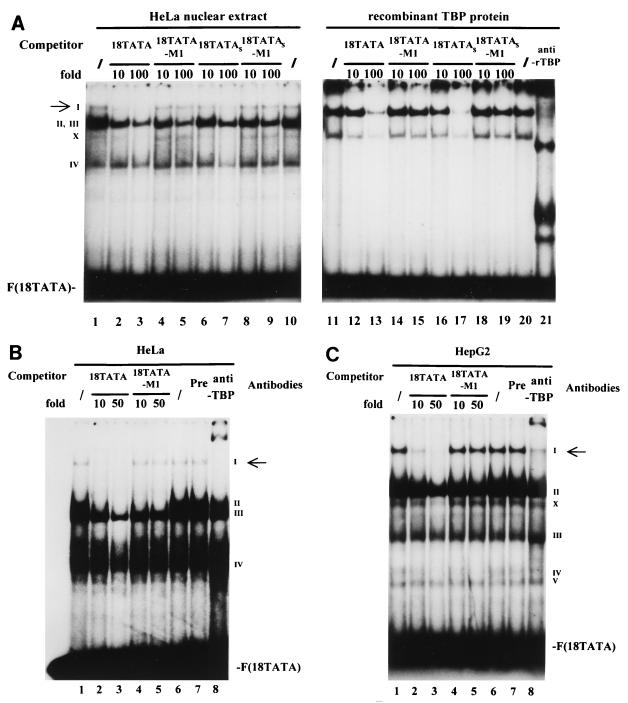
FIG. 6.
Binding of TBP to the TATA box of the HPV-18 URR. (A) Formation of the specific TBP-TATA box complex I. 32P-labeled oligonucleotide 18TATA was used in EMSAs with HeLa nuclear extracts (lanes 1 to 10) or bacterial rTBP (lanes 11 to 21). Binding reactions were carried out with HeLa cell nuclear extracts in the absence (/) (lanes 1 and 10) or presence (lanes 2 to 9) of unlabeled competitor oligonucleotides. Unlabeled competitor oligonucleotides 18TATA, 18TATA-M1, 18TATAS, and 18TATAS-M1 were each used in 10- and 100-fold molar excesses, as indicated above the lanes. Binding reactions were carried out with bacterial rTBP in the absence (/) (lanes 11 and 20) or presence (lanes 12 to 19) of unlabeled competitor oligonucleotides. Unlabeled competitor oligonucleotides were used in the same manner as for the competition experiments with HeLa cell nuclear extracts. A specific antibody against bacterial rTBP (anti-rTBP) was included in the binding reaction shown in lane 21. A specific DNA-protein complex is indicated by a I (marked by an arrow); numerals II to IV indicate nonspecific DNA-protein complexes. The complex marked by an X is likely caused by binding of partially degraded TBP to the probe. F(18TATA), free probe 18TATA. EMSAs were resolved on a 6.5% polyacrylamide gel. (B) Anti-human TBP antibody specifically recognizes complex I in HeLa cells. Binding reactions with 32P-labeled 18TATA were carried out with HeLa cell nuclear extracts in the absence (/) (lanes 1 and 6) and presence (lanes 2 to 5) of unlabeled competitor. Unlabeled competitor oligonucleotides 18TATA and 18TATA-M1 were used in a 10- or 50-fold molar excess, as indicated above the lanes. A specific antibody against human TBP protein (1 μl) was included in the binding reaction shown in lane 8 (anti-TBP). In lane 7, the binding reaction mixture included 1 μl of preimmune serum (Pre). A specific DNA-protein complex is indicated by the I (marked by an arrow); numerals II to IV indicate nonspecific DNA-protein complexes. F(18TATA), free probe 18TATA. (C) Anti-human TBP antibody specifically recognizes complex I in HepG2 cells. Essentially the same binding reactions as described for panel B were carried out with nuclear extracts from HepG2 cells. A specific DNA-protein complex is indicated by the I (marked by an arrow); numerals II to V indicate nonspecific DNA-protein complexes. The complex marked by an X is likely caused by binding of partially degraded TBP to the probe. F(18TATA), free probe 18TATA.

