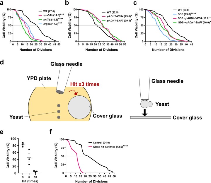
Extended Data Fig. 5. plasma membrane damage limits replicative lifespan in budding yeast.
(a-c) Yeast replicative life span was measured with another biological replicate, and results were consistent with Fig. 2 (e-g). Cell number: (a) Wild type (n = 24), vps34Δ (n = 24), snf7Δ (n = 24), erg2Δ (n = 24). (b) Wild type (n = 48), VPS4 (n = 48), SNF7 (n = 48). (c) Wild type (n = 24), SDS (n = 24), SDS+VPS4 (n = 24), SDS+SNF7 (n = 24). P value: *<0.05, **<0.01, ****<0.001 by two-sided Wilcoxon Rank-sum test. Exact P value: (a) WT vs. vps34Δ: 0.0005, WT vs. snf7Δ: <0.0001, WT vs. erg2Δ: 0.0008. (b) WT vs. VPS4: 0.0209, WT vs. SNF7: 0.0465. (c) WT vs. SDS: <0.0001, WT vs. SDS+VPS4: 0.0473, WT vs. SDS+SNF7: 0.0158. (d) A schematic image of mechanical damage induced by a glass needle and a glass coverslip. (e) The viability of the yeast cells was measured. Mean(SD) of 3 biological replicates. n = 40. (f) WT yeast cells were subjected to the replicative life span measurement with or without mechanical damage. Median values are shown. Cell number: Wild type (n = 24), Glass (n = 24). P value: ****<0.001, exact value: <0.0001, by two-sided Wilcoxon Rank-sum test.

