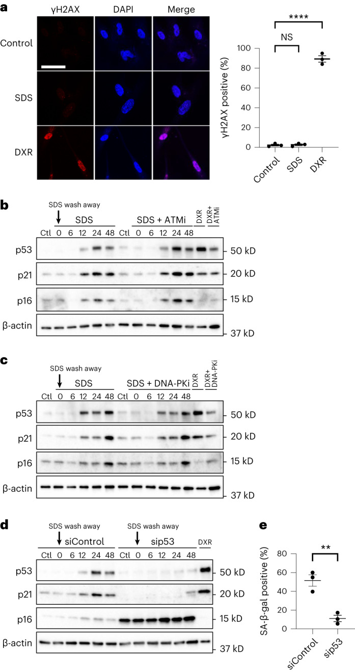
Fig. 5. PMD-dependent senescence requires p53.

a–e, WI-38 cells were treated with 0.007% SDS or 250 nM DXR combined with indicated additional treatment for 24 h, washed and released into fresh medium. a, γH2AX staining. WI-38 cells at 24 h after SDS or DXR wash away and untreated cells (control) were stained with γH2AX (red) and with DAPI (blue). Scale bar, 50 μm. Graphs show quantification of γH2AX-positive cells (n > 100 cells). ****P < 0.001, control versus SDS: 0.9896 and control versus DXR: <0.0001, by one-way ANOVA with Dunnettʼs test. b–d, Western blotting using cell lysates of WI-38 cells treated with SDS with or without ATM inhibitor KU-55944 (10 μM) (b), DNA-PK inhibitor (10 μM) (c) and p53 siRNA (d). e, SA-β-gal-positive cells were counted on 10 d after wash away. WI-38 cells were treated with p53 siRNA (sip53) or control scramble siRNA (siControl). **P < 0.01, exact value: 0.0091, by two-tailed unpaired Student’s t-test. Data in a and e are presented as mean (horizontal bars) ± s.d. (whiskers) of three biological replicates. NS, not significant.
