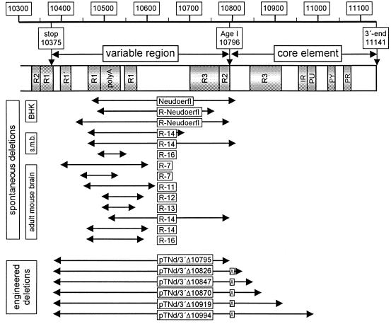
FIG. 1.
Schematic representation of the 3′ NCR of TBE virus Neudoerfl. Numbers correspond to the full-length genomic sequence (GenBank accession no. U27495). Shaded areas depict sequence elements that were described previously (15, 31): R1, R2, R3, direct repeats; polyA, internal poly(A) sequence, here 49 residues long, as it is in the infectious cDNA clone of TBE virus; IR, inverted repeat; PU, homopurine box; PY, homopyrimidine box; PR, pyrimidine-rich box. The positions of spontaneous 3′-NCR deletions observed during cell culture growth (BHK), in suckling-mouse brain stock solutions (s.m.b.), and in virus isolated from adult mouse brains are aligned below the schematic. For the exact positions of the deletion boundaries, refer to Tables 1 and 2. The bottom section shows the engineered deletions which extend from nucleotide 10378 to the positions as given in the corresponding plasmid name. A boxed A represents the 6-nucleotide AgeI recognition sequence which was retained in the construction procedure.

