
Fig. 4.
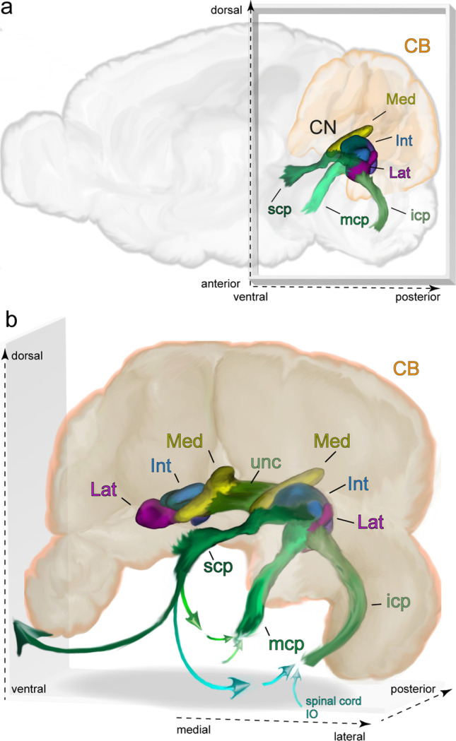
Overview of the CN of the mouse and the fiber bundles connecting them to the rest of the brain. a The location of CN and fiber bundles in a sagittal schematic of the mouse brain. b A depiction of the CN within the cerebellum. Arrows indicate the primary directions of axonal projections within the bundles. Dark green and light blue connections via the superior cerebellar peduncle indicate ascending and descending connections. The two arrows feeding into the icp and mcp indicate a combination of inputs arriving from ascending pathways (e.g., from the spinal cord or inferior olive) and descending ones (e.g. via the basal pontine nuclei). Note that, for clarity, the brainstem is not shown. Abbreviations: CB, cerebellum; Med, medial nucleus; Int, interposed nucleus; Lat, lateral nucleus; scp, superior cerebellar peduncle; mcp, middle cerebellar peduncle; icp, inferior cerebellar peduncle; unc, uncinate fibers; IO, inferior olive