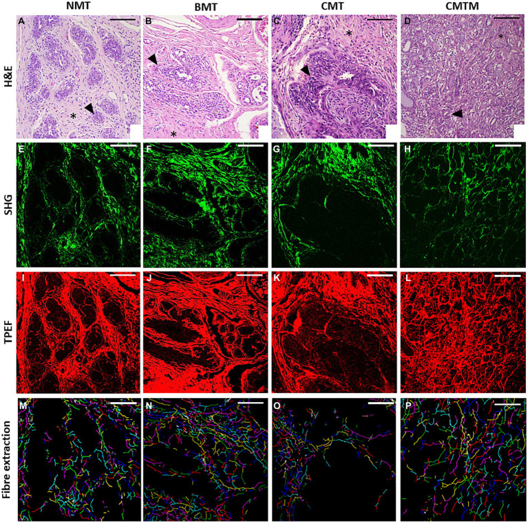
Figure 1.
Acquired images (x20). The optical microscopy H&E (A–D), second harmonic generation microscopy (E–H), two-photon excited fluorescence microscopy (I–L), and the extracted collagen fibres with each fibre in a random colour (M–P) for the canine normal mammary tissue (A,E,I,M), benign mixed tumour (B,F,J,N), carcinoma in the mixed tumour without metastasis (C,G,K,O) and carcinoma in mixed tumour with metastasis (D,H,L,P). HE asterisks: stroma. HE arrowhead: epithelial cells. The scale bar is 100 μm.

