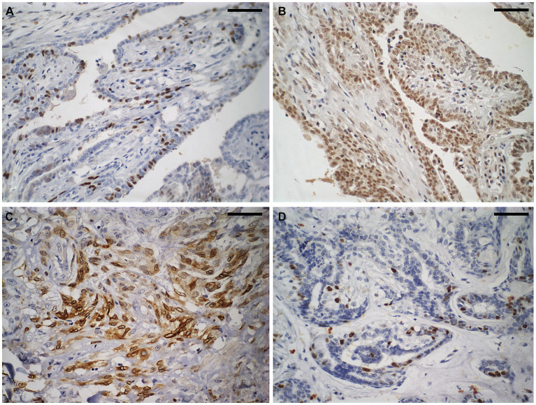
Figure 2.
Immunostaining to evaluate the molecular subtype of carcinomas in mixed tumours of the canine mammary gland. Shown here is a carcinoma in a mixed tumour without lymph node metastasis. (A) Strong nuclear immunostaining for oestrogen receptor in approximately 25% of neoplastic cells. Diaminobenzidine chromogen, haematoxylin counterstaining. (B) Strong nuclear immunostaining for progesterone receptor (brown areas) in more than 75% of neoplastic cells. Diaminobenzidine chromogen, haematoxylin counterstaining. (C) Strong cytoplasmic immunostaining for cyclooxygenase-2 in approximately 60% of neoplastic cells. Diaminobenzidine chromogen, haematoxylin counterstaining. (D) Strong nuclear immunostaining for Ki67 in approximately 10% of neoplastic cells. Diaminobenzidine chromogen, haematoxylin counterstaining. The scale bar is 50 μm.

