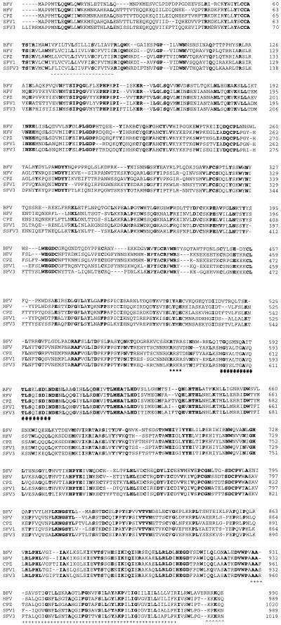
FIG. 4.
Alignment of foamy virus Env proteins. Identical amino acids conserved among the foamy viruses are shown in bold. Putative functional domains are shown; the putative leader peptide is underlined with dashes, the proposed SU-TM cleavage site is marked by asterisks, the cell fusion domain is marked by number signs, the transmembrane segment is marked by plus signs, and the basic cytoplasmic tail is marked by carets. The amino acid positions of the Env proteins are to the right of the sequences.

