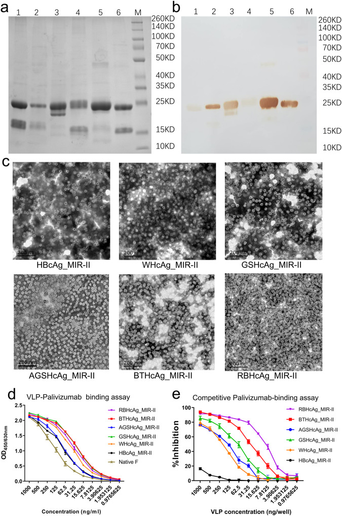
Fig. 2. Recombinant expression, characterization, and bioactivity evaluation of the designed chimeric VLPs presenting RSV F epitope II by using different species of HcAg as the scaffolds.
The designed chimeric VLPs were expressed by transfecting the hansenula polymorpha using a system developed by our laboratory. a, b SDS-PAGE (a) and Western-blot using palivizumab (b) for the designed chimeric VLPs, respectively. Lane 1: BTHcAg_MIR-II, Lane 2: AGSHcAg_MIR-II, Lane 3: WHcAg_MIR-II, Lane 4: HBcAg_MIR-II, Lane 5: GSHcAg_MIR-II, Lane 6: RBHcAg_MIR-II, Lane M: Protein molecular weight marker. c TEM microscopic images of the designed chimeric VLPs (scale bar 200 nm). d Concentration-dependent binding abilities of the designed chimeric VLPs to palivizumab tested using ELISA, which were compared with that of the native RSV F protein. Triplicate measurements were performed, and data are presented as mean ± SEM. e Competitive palivizumab-binding abilities of the designed chimeric VLPs in contrast with the native RSV F protein tested using ELISA. In this assay, the concentration of the native RSV F protein was 0.5 μg/ml (50 ng/well), and the VLP samples were serially diluted to different concentrations. Triplicate measurements were performed, and data are presented as mean ± SEM.

