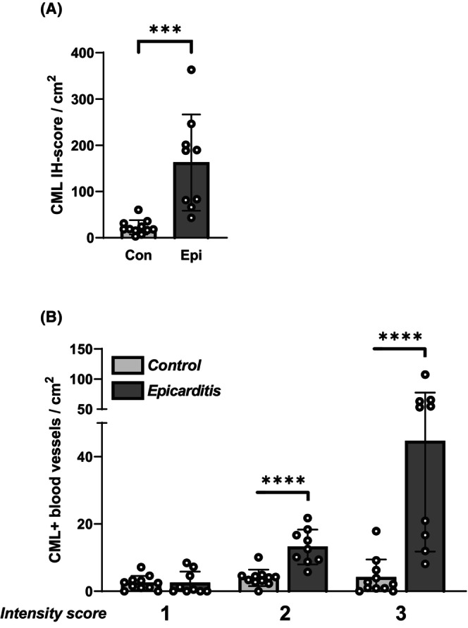
FIGURE 2.

Quantification of N(e)‐(carboxymethyl)lysine (CML) in the intramyocardial vasculature. Shown are (A): the CML‐immunohistochemical (IH) score, as well as (B): the numbers of intramyocardial blood vessels with weak‐ (intensity score 1); moderate‐ (intensity score 2); and strong‐ (intensity score 3) CML staining. ***p = .0001, ****p < .0001.
