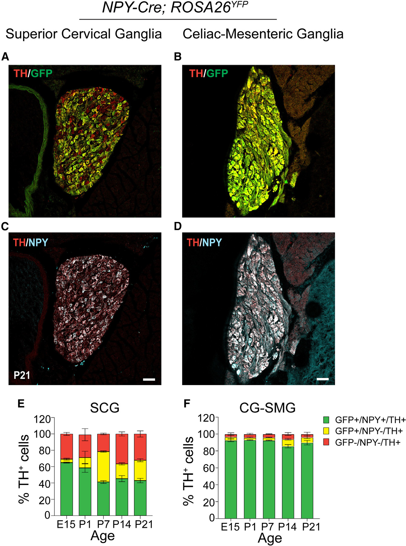
Figure 1. Differential NPY expression in paravertebral and prevertebral sympathetic neurons.

(A–D) Immunostaining for TH, NPY, and GFP in superior cervical ganglia (SCGs; paravertebral) (A and C) and celiac-superior mesenteric ganglia (CG-SMGs; prevertebral) (B and D) in NPY-Cre:ROSA26EYFP reporter mice.
(E and F) Percentage of NPY-expressing noradrenergic neurons in SCGs (E) and CG-SMGs (F). NPY and TH are co-expressed (green bar) in ~65% of SCG neurons at E15 and down-regulated to ~43% at P21. NPY and TH are co-expressed in ~90% of CG-SMG neurons throughout development. Neurons that expressed NPY earlier but no longer do so at the time of examination are represented by the yellow bars. Sympathetic neurons that never express NPY are indicated by red bars. n = 3 mice per genotype for each time point. Scale bars, 50 μm.
