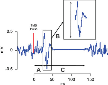
Figure 1. Features of the MEP.

The motor threshold reflects the minimum intensity that elicits a small MEP in 50% of trials and can be probed at rest and during tonic muscle contraction. Motor thresholds are thought to rely on the excitability of cortico‐cortical axons since voltage‐gated sodium channel‐blocking drugs increase thresholds (Ziemann, 2013). It is important to note that motor thresholds vary tremendously across individuals and do not represent a static measure. For instance, differences in scalp‐to‐cortex distance and genetic factors may explain a lot of the variance in the motor threshold between individuals. A, MEP latency provides information about the conduction time for the neural responses triggered by TMS to reach the targeted muscle, including the time for excitation of cortical neurons, conduction of the pyramidal tract and summation of descending volleys at the spinal level, and conduction time of peripheral motor neurons. The latency, therefore, can be influenced by both the state of the cortical and spinal motor neuron pool and certain stimulation parameters. For instance, MEP latencies are shortened with voluntary contraction as this action reactivates spinal motoneurons and in turn lowers their firing threshold. B, MEP size can be measured either by measuring peak‐to‐peak amplitude or the area under the curve of the rectified MEP. With either measure, it is possible to test TMS recruitment curves that establish the input–output relationship between increasing TMS intensity and resulting MEP size. While measuring MEP size as peak‐to‐peak amplitude is more commonly used, this metric is only valid when there is no occurrence of polyphasic oscillations. For example, MEPs elicited for non‐hand muscles tend to be more polyphasic (Groppa et al., 2012), as well as those recorded from various patent populations, including patients with the amyotrophic lateral disease (Kohara et al., 1999), myoclonus dystonia (Van Der Salm et al., 2009), multiple sclerosis (Kukowski, 1993) and stroke (Brum et al., 2016). C, silent period represents a period of reduced electrical activity that follows the MEP when elicited during voluntary contraction. The duration depends on the stimulus intensity and is influenced by both intracortical and spinal mechanisms. The initial portion has been suggested to be due to a contribution from spinal mechanisms involving changes in motoneuron excitability and recurrent inhibition (Fuhr et al., 1991). The latter part has been linked to cortical inhibition mediated by GABAB (Ziemann et al., 2004).
