Figure 1.

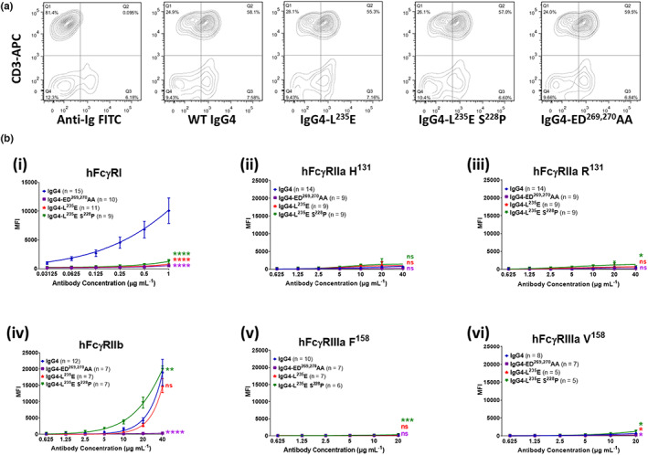
CD28‐ and FcγR‐binding properties of anti‐CD28 wild‐type and mutant antibodies. (a) Anti‐CD28 mAb mutants, detected using anti‐human Ig‐FITC (x axis), binding to human PBMCs costained with mouse anti‐CD3‐APC, as a marker for T cells (y axis). Data are representative of three independent experiments. (b) Binding of anti‐CD28 IgG4 mAbs to human FcγRs as detected by flow cytometry. (i) FcγRI binding was determined by incubating cells with monomeric IgG mutants (0.03–1 μg mL−1) and detected using Alexa Fluor 647–conjugated F(ab′)2 fragments of anti‐human IgG F(ab′)2. (ii–vi), the binding of complexed CD28 mAbs to low‐affinity receptors FcγRIIa, FcγRIIb and FcγRIIIa was performed by preincubating antibodies (20 μg mL−1) with Alexa Fluor 647–conjugated F(ab′)2 fragments of anti‐human IgG F(ab′)2 (10 μg mL−1), then titrating these complexes and incubating with the FcR cells. Analysis was performed with FlowJo. Data are the mean ± standard error of the mean for 5–15 experiments analyzed by two‐way ANOVA with Dunnett's multiple comparisons test, comparing the main column effect with unmodified IgG4; ns, not significant, *P ≤ 0.05, **P ≤ 0.01, ***P ≤ 0.001, ****P ≤ 0.0001. APC, allophycocyanin; FcγR, Fcγ receptor; FITC, fluorescein isothiocyanate; Ig, immunoglobulin; mAb, monoclonal antibody; MFI, mean fluorescent intensity; PBMC, peripheral blood mononuclear cell; WT, wild‐type.
