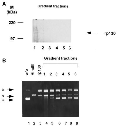
FIG. 6.
Single-step purification of rp130 and direct association with enzyme activity. (A) Recombinant-infected cells were lysed and layered on a 5 to 20% sucrose gradient. Fractions were collected, separated by SDS-PAGE (8% gel) and transferred to a polyvinylidene difluoride membrane. Detection was performed with an anti-p130 antibody. Lane 1, fraction 1; lane 2, fraction 2; lane 3, fraction 3; lane 4, fraction 4; lane 5, fraction 5; lane 6, fraction 6. Molecular mass markers (M) are shown on the left; the position of rp130 is indicated by the arrow on the right. (B) Nuclease activity was assayed with an aliquot of each fraction. Lanes: 1, pON205 alone; 2, pON205 treated with HindIII; 3, pON205 plus extract containing rp130; 4, plasmid plus protein of fraction 1; 5, incubation with an aliquot of fraction 2; 6, incubation with an aliquot of fraction 3; 7, incubation with an aliquot of fraction 3; 8, incubation with an aliquot of fraction 4; 8, incubation with an aliquot of fraction 5; 9, incubation with an aliquot of fraction 6. The arrows indicate three different plasmid forms: open circular (a), linear (b), and supercoiled (c).

