Figure 1.

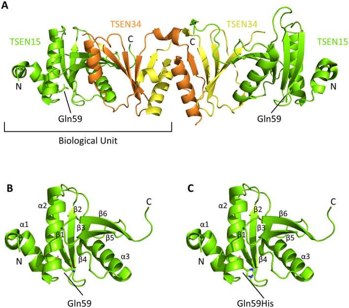
TSEN15 protein structure. A, Crystal structure of the TSEN15 (green)–TSEN34 (yellow and orange) heterodimer (Protein Data Bank, identification no. 6Z9U). The position of the Gln59 residue is highlighted, and the amino acid side chain is displayed. B, Monomeric crystal structure of TSEN15 (Protein Data Bank, identification no. 2GW6), showing numbering of the α‐α‐β‐β‐β‐β‐α‐β‐β fold (α1–3 and β1–6). Gln59 is labeled, and the side chain is displayed. C, TSEN15 structure, shown as in B, following in silico mutagenesis to predict the conformation of Gln59‐His (labeled). In B and C, red shows oxygen atoms and blue shows nitrogen atoms. The PyMOL Molecular Graphics System was used to view structures and to perform mutagenesis.
