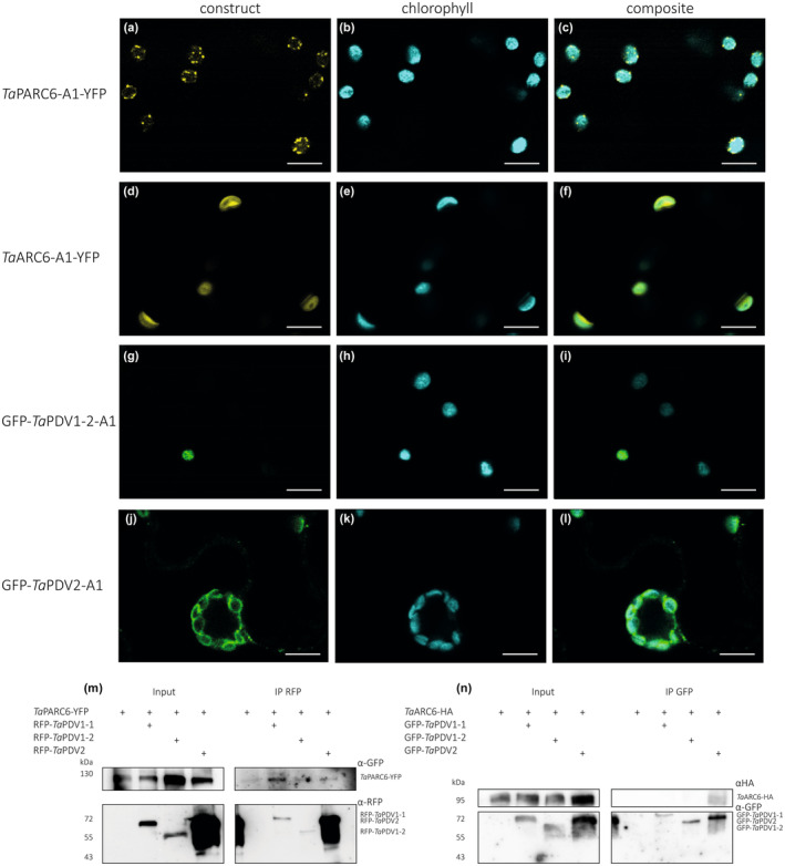
Fig. 7.

Localisation and co‐immunoprecipitation assays of wheat TaPARC6, TaARC6 and TaPDV isoforms. (a–l) Images of transiently expressed AtUbi:TaPARC6‐A1‐YFP, CaMV35S:TaARC6‐A1‐YFP, CaMV35S:GFP‐TaPDV1‐2‐A1 and CaMV35S:GFP‐TaPDV2‐A1 in Nicotiana benthamiana epidermal cells. Images were acquired using confocal laser scanning microscopy. The yellow fluorescent protein (YFP) and green fluorescent protein (GFP) fluorescence are shown in yellow and green, while chlorophyll autofluorescence is shown in cyan. Bar, 10 μm. (m) Immunoprecipitation (IP) assay using anti‐red fluorescent protein (RFP) beads for interactions between TaPARC6‐YFP and RFP‐TaPDV1‐1, RFP‐TaPDV1‐2 and RFP‐TaPDV2, transiently co‐expressed in Nicotiana leaves. Immunoblots RFP and GFP antibodies were used to detect the proteins. (n) Immunoprecipitation (IP) assay using anti‐GFP beads for TaARC6‐HA and GFP‐TaPDV1‐1, GFP‐TaPDV1‐2 and GFP‐TaPDV2, transiently co‐expressed in Nicotiana leaves. Immunoblots with HA (hemagglutinin)‐tag and GFP antibodies were used to detect the proteins.
