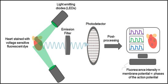
Figure 1. Principle of optical mapping.

The heart stained with a voltage‐sensitive fluorescent dye is illuminated with a specific wavelength of light (e.g. 532 nm for the excitation of Di‐4‐ANEPPS). The dye emits voltage‐dependent fluorescent light, which is first filtered using an emission filter and then captured on a high‐resolution photodetector. The recorded intensity on the photodetector is post‐processed to obtain optical action potential signals.
