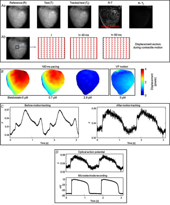
Figure 3. 2D motion tracking.

A, motion tracking algorithm computes the geometrical transformation between test (T) images and a reference (R) image and aligns the test images with the reference image (image registration). a, the difference image between the test and the reference image shows significant motion artifacts before motion tracking (R − T). Motion artifacts are significantly reduced in the difference image after motion tracking (R − Tt). b, displacement vectors computed via the motion tracking algorithm at three different time points (40 ms intervals) during cardiac contraction. B, motion amplitude maps (in pixels) computed using 2D motion tracking during ventricular pacing and fibrillation, showing relatively less motion in VF than during ventricular pacing. Panel modified from our previous study (Kappadan et al., 2020). C, example of motion tracking in frog heart, where blebbistatin does not work. Motion tracking significantly reduced the distortion of optical action potentials (motion artifacts). Signals are spatially averaged from 3 × 3 pixels. D, validation of motion tracking: comparison of motion tracked optical action potential and a microelectrode action potential recording of a frog heart at 1.5 s period showing good agreement between the signals.
