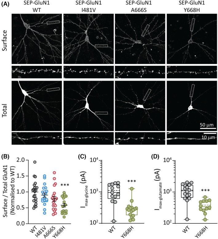
FIGURE 2.

GluN1 variant surface expression. (A) Rat hippocampal neurons transfected with plasmids encoding superecliptic pHluorin (SEP)‐GluN1 (wild‐type [WT]) and the three variant subunits SEP‐GluN1(I481V), SEP‐GluN1(A666S), and SEP‐GluN1(Y668H). Representative images are shown of the surface and total SEP‐GluN1 in a neuron from each group, together with expanded views of the boxed regions, shown below. (B) Quantification of surface expression of the surface/total GluN1 ratio normalized to the value of control neurons expressing SEP‐GluN1 WT. Data are presented as mean ± SEM (WT, n = 20 neurons; I481V, n = 20; A666S, n = 20; Y668H, n = 20; from three independent cultures). (C) Quantification of peak whole‐cell currents mediated by the indicated receptors in response to saturating concentrations of glycine (and half‐maximal effective concentration [EC50] of glutamate). (D) Quantification of peak whole‐cell currents in response to saturating concentrations of glutamate (and an EC50 concentration of glycine). ***p < .001.
