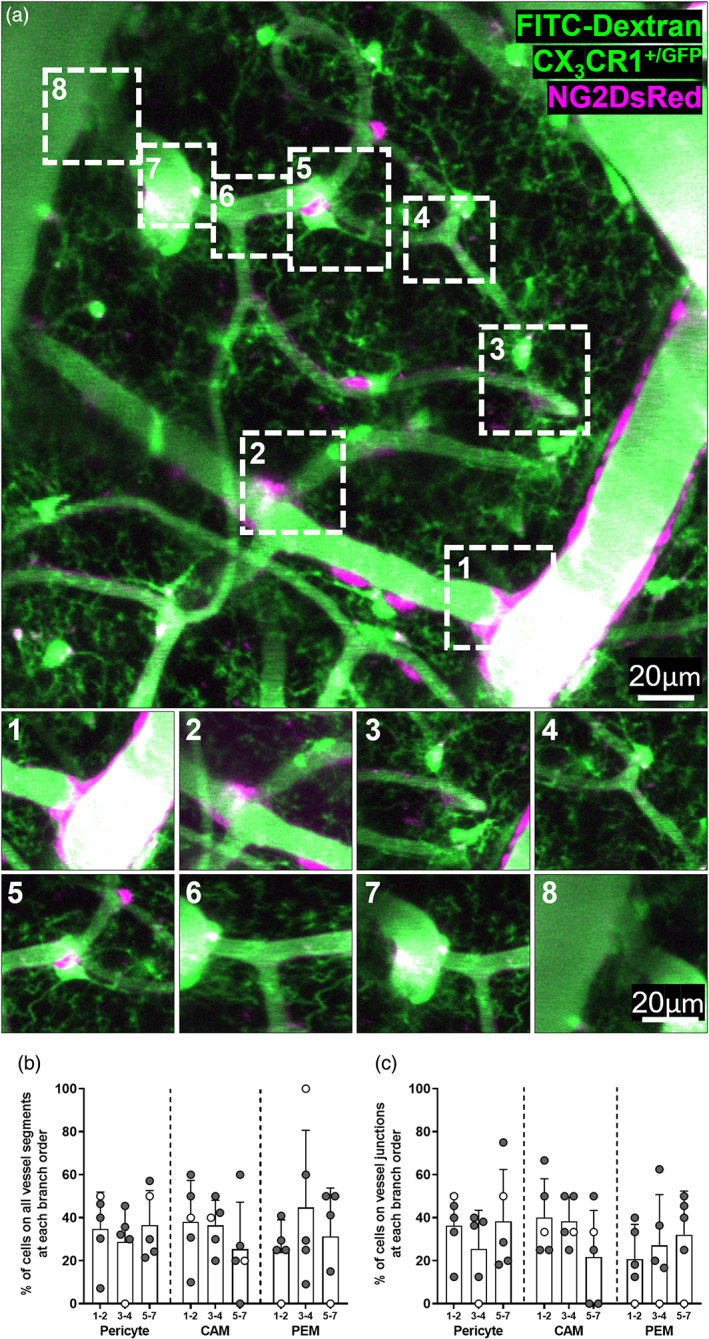
FIGURE 2.

CAM, PEM and pericytes are present at all branch orders of capillaries. (a) Representative image stack (20–75 μm depth, average intensity projection) of FITC dextran filled vessels in the somatosensory cortex of an NG2DsRed × CX3CR1+/GFP mouse imaged in vivo using 2PLSM. Pericytes are labeled magenta, and microglia and vessels are green. Insets correspond to boxes labeled 1–8 in main panel. (1) Penetrating arteriole (0th order) branching off to form a capillary (first order). (2–7) Higher order capillaries branching. (8) Seventh order capillary converging on the ascending venule. (b, c) Percentage of total CAM, PEM and pericytes that are located (b) at different branch orders of the vascular tree, and (c) at vessel junctions at different branch orders of the vascular tree (n = 5, four male and one female). For all graphs, gray circles represent males and white circles represent females. Data presented as mean ± SD.
