FIGURE 3.

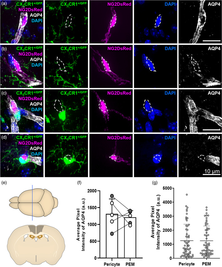
Microglia can interact with pericytes with or without AQP4‐positive astrocyte endfeet. (a) Representative example of a pericyte (magenta) with no associated PEM exhibiting strong labeling by AQP4 (white). (b) Representative example of a pericyte (magenta) that is not associated with a PEM, but lacking AQP4 (white) labeling. (c) Representative example of a pericyte (magenta) associated with a PEM (green). The pericyte is exhibiting strong labeling by AQP4 (white). (d) Representative example of a pericyte (magenta) associated with a PEM (green). The pericyte is lacking AQP4 (white) labeling. (e) Schematic of region analyzed in 12‐week‐old NG2DsRed × CX3CR1+/GFP mice (~Bregma −1.10 mm [Allen Reference Atlas, n.d.]). (f) Quantification of AQP4 fluorescence intensity in regions overlaying pericytes with, or without, an associated PEM (n = 5, two males, three females). Data compared with a paired parametric t‐test. (g) AQP4 fluorescence intensity around individual pericytes with, or without, an associated PEM (n = 5, two males, three females). This data was used to derive the averages in (f). Data are presented as mean ± SD. For all images: NG2DsRed‐positive pericytes (magenta), CX3CR1+/GFP‐positive microglia (green), AQP4‐labeled astrocyte endfeet (white) and DAPI‐labeled nuclei (blue) are shown. Images showing each fluorescent channel alone are to the right of the main image.
