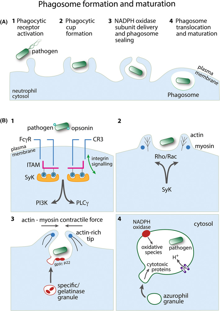
FIGURE 1.

Phagosome formation and maturation. (A) Overview of the major steps in neutrophil phagosome formation following pathogen detection. Events 1–4 are depicted in further detail in panel B. (B) 1: An opsonized pathogen engages Fc receptors (FcγR) or complement receptors (e.g., CR3) to initiate phagocytosis. Both FcγR and CR3 can employ immunoreceptor signaling pathways: SH2‐domain‐bearing proteins (e.g., Syk) associate with phosphorylated ITAM, signaling downstream through phosphatidylinositol 3‐kinase (PI3K) and/or phospholipase Cγ (PLCγ). CR3 also employs independent inside‐out and outside‐in integrin signaling pathways. 2: Phagocytic receptor signaling induces regulation of the actin cytoskeleton via Rac and/or Rho. Myosin motor control of actin rearrangement drives extending pseudopod protrusions from the plasma membrane to form the phagocytic cup around the pathogen. 3: Cytosolic specific/gelatinase granules deliver proteins to the membrane of the forming phagosome, for example, the membrane‐bound subunits of NADPH oxidase, gp91phox (NOX2), and p22phox. Actin polymerization at the pseudopod tips facilitates membrane sealing to complete the phagocytic vacuole around the pathogen. 4: The formed pathogen‐containing phagosome translocates toward the granule‐rich centriole within the neutrophil cytosol. NADPH oxidase generates antimicrobial reactive oxygen species inside the phagosome. The negative charge generated by this process is compensated by an influx of protons. Cytosolic azurophil granules, containing cytotoxic proteins, for example, elastase, fuse with the phagosome membrane to deliver their contents to the lumen of the phagosome
