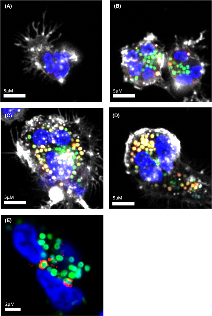
FIGURE 2.

Maturation of phagosomes formed by human peripheral blood neutrophils ingesting Staphylococcus aureus. (A) Neutrophil that has not encountered bacteria (Blue: Nucleus labeled with Hoescht, White: Actin labeled with Alexa Fluor(AF)647‐phalloidin). (B) Early ingestion of S. aureus (Green: AF488‐labeled S. aureus bioparticles), with limited change in phagosomal pH as assessed by co‐localized lysotracker signal (Red: Lyso Tracker Red DND‐99, Yellow: co‐localization with AF488 S. aureus). (C): Mid‐maturation with mixture of low pH (mature) and high pH (immature) phagosomes. (D): Late maturation with the majority of phagosomes demonstrating low pH. (E) Early endosomal antigen 1 (EEA1) staining on the surface of S. aureus containing phagosomes. Note the penumbral rather than co‐localized signature indicating membrane distribution (Red: EEA1, mouse anti‐human EEA1 with secondary anti‐mouse AF568 phalloidin staining omitted for clarity)
