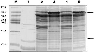
FIG. 3.
Coomassie blue-stained SDS-PAGE (15% polyacrylamide) gel of expressed recombinant fiber knob proteins. Lanes: M, molecular mass markers (the sizes of the markers are indicated on the left in kilodaltons); 1, uninduced E. coli M15 cells (transformed with pQE); 2, fiber knob protein FK17GCE (sample boiled prior to loading); 3, fiber knob protein FK17GCE (sample not boiled); 4, fiber knob protein FK17S (sample boiled prior to loading); 5, fiber knob protein FK17S (sample not boiled). The arrows indicate the positions of the monomeric and trimeric fiber knob proteins.

