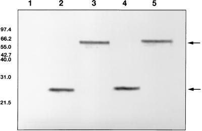
FIG. 4.
Western blot (of 15% polyacrylamide gel). Lanes: 1, uninduced E. coli M15 cells (transformed with pQE); 2, fiber knob protein FK17GCE (sample boiled prior to loading); 3, fiber knob protein FK17GCE (sample not boiled); 4, fiber knob protein FK17S (sample boiled prior to loading); 5, fiber knob protein FK17S (sample not boiled). The sizes of the markers are indicated on the left in kilodaltons. The arrows indicate the positions of the monomeric and trimeric fiber knob proteins.

