
Figure 2.
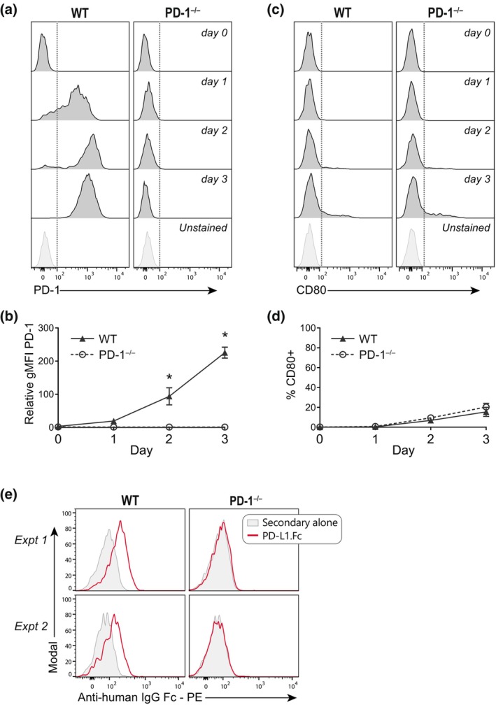
PD‐L1.Fc does not inhibit CD8+ T cells via specific surface receptor interactions. (a–d) Programmed death receptor 1 (PD‐1) and CD80 expression on wild‐type (WT) and PD‐1−/− CD8+ T cells following anti‐CD3 + anti‐CD28 stimulation as in Figure 1. (a) Representative histograms of PD‐1 expression over time. (b) Summary of PD‐1 geometric mean fluorescence intensity relative to unstained in WT (triangles) and PD‐1−/− (open circles) T cells in four independent experiments (mean ± standard error of the mean). (c) Representative histograms of CD80 expression over time. (d) Summary of % CD80+ T cells in WT (triangles) and PD‐1−/− (open circles) T cells in three independent experiments (mean ± standard error of the mean). (e) Detection of PD‐L1.Fc binding to WT versus PD‐1−/− CD8+ T cells day 2 after activation with anti‐CD3 + anti‐CD28 (two independent experiments). Unpaired t‐tests were performed in b and d comparing WT versus PD‐1−/− T cells. *P < 0.05. Ig, immunoglobulin; PD‐L1, PD‐1 ligand 1; PD‐L1.Fc, PD‐L1 coupled to the Fc portion of IgG1; PE, phycoerythrin.