
Figure 3.
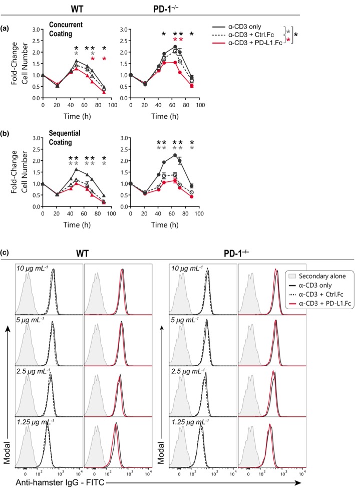
Protein concentration does not account for PD‐L1.Fc inhibition. (a, b) Wild‐type (WT) and PD‐1−/− CD8+ T cells were stimulated on plates coated with anti‐CD3 only (black), anti‐CD3 + Ctrl.Fc (black, dashed‐open) or anti‐CD3 + PD‐L1.Fc (red), with anti‐CD28 costimulation and endogenous interleukin (IL)‐2 neutralized with anti‐IL‐2 monoclonal antibody (mAb). Analysis of cell number over time (mean ± standard error of the mean, of triplicate wells) of cells stimulated on plates coated concurrently with anti‐CD3 mAb and Fc (a, representative of two independent experiments), or coated overnight (4°C) with anti‐CD3 mAb followed by 4 h (37°C) with Fc (b, one experiment). (c) Detection of anti‐CD3 mAb (1.25–10 μg mL−1) binding on naïve WT or PD‐1−/− CD8+ T cells with co‐incubation of anti‐CD3 alone or with equivalent concentrations of Ctrl.Fc or PD‐L1.Fc (1 experiment). Unpaired t‐tests were performed comparing conditions at each timepoint in a and b. *P < 0.05. PD‐1, programmed death receptor 1; PD‐L1, PD‐1 ligand 1; PD‐L1.Fc, PD‐L1 coupled to the Fc portion of IgG1.