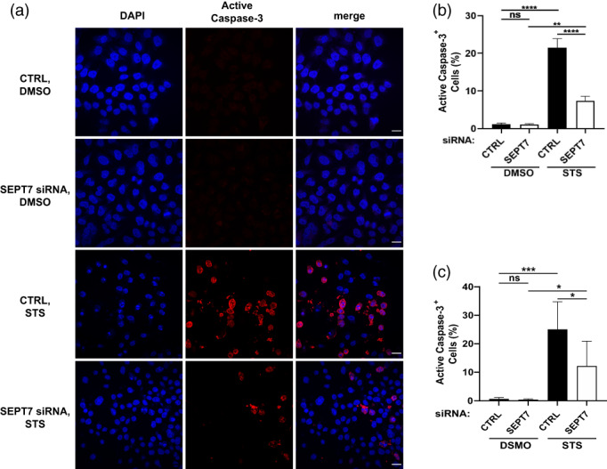
FIGURE 4.

SEPT7 is required for the activation of caspase‐3. (a) Control and SEPT7‐depleted cells were treated with DMSO or STS, and stained with cleaved anti‐caspase‐3 antibody and DAPI. Representative confocal microscopy images showing cleaved caspase‐3 in red and DAPI in blue. Scale bar = 100 μm. (b) The percentage of cleaved caspase‐3‐stained cells was calculated in Fiji by dividing the number of cells that displayed cleaved caspase‐3 staining by the total number of cells, then multiplying by 100. Each bar represents the mean % ± SEM from three independent experiments (a minimum of 1,000 cells were counted per condition separated in three independent experiments). **p < .01, ****p < .0001 by two‐way ANOVA. (c) HeLa cells (CTRL or SEPT7 siRNA transfected) were treated with 1 μM staurosporine for 5 hr, followed by staining of active caspase‐3 and analyzed by flow cytometry (a minimum of cells were counted per condition separated in three independent experiments). *p < 0.05, ***p < 0.001 by two‐way ANOVA
