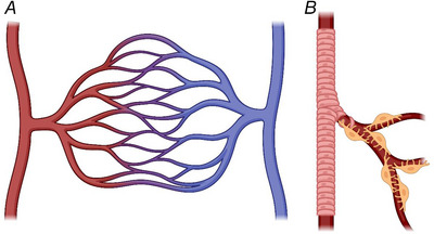
Figure 1. Typical arrangement of the microvasculature.

A, a simple diagram showing the transit and direction of blood flow from arterioles (red, on the left) which progress into a larger capillary bed before ending in venular output (blue, on the right). B, cartoon illustrating the arrangement of smooth muscle cells around an arteriole (pink), in contrast to the pericyte‐covered capillary bed with thin pericyte processes extending along and around the vessel (pericytes shown in yellow). Produced using BioRender.
