Fig. 1.
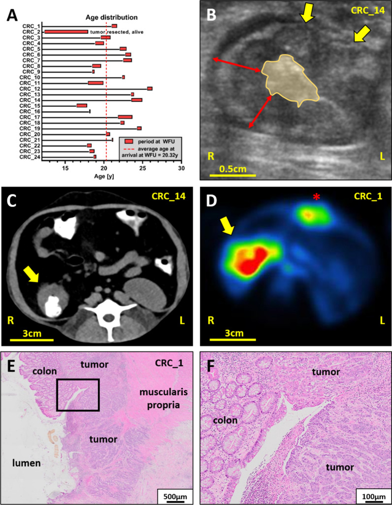
Colorectal cancer in rhesus macaques—age, diagnostic imaging and histopathology. A Age distribution of CRC-bearing rhesus macaques at the age of arrival at WFUSM and upon elective or clinical endpoints. B US imaging illustrates the loss of colonic wall architecture and replacement by nodular lesions (yellow arrows), the circumferential thickening of the colonic wall ranging from 0.5 to 1 cm (red arrows) and a constricted lumen (yellow area). C Iodine-based oral-contrast improves the accuracy of CTs by delineating the constricted lumen and colonic wall thickening (yellow arrow) at the ileocecal junction. D 18F-FDG-PET imaging highlights a circumferential region of high metabolic activity at the proximal colon (yellow arrow) and high metabolic activity at the healing laparotomy site (red asterisk). E + F H&E sections used for staging and grading illustrate the effaced colonic architecture and the penetration of both the circumferential and longitudinal muscle layers of the muscularis propria by the CRC. Note, the black rectangle in (E) highlights the area depicted with higher magnification in (F)

