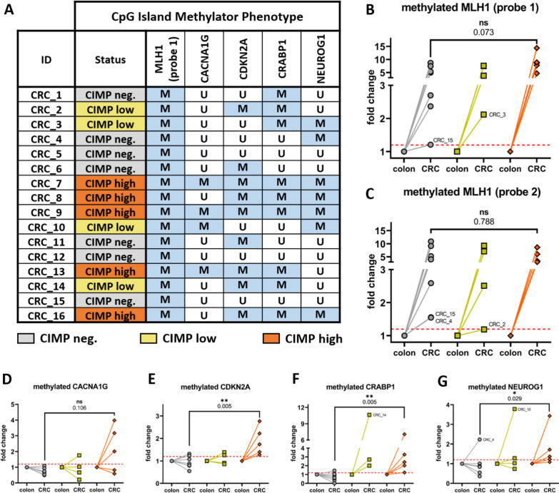
Fig. 5.
MLH1 hypermethylation occurs in all examined rhesus CRCs and is independent of the more global CIMP status. A marker was considered hypermethylated in CRC upon an increase of 20% in DNA methylation (red dotted line) in comparison to paired healthy colon as reference. A As a result, 5/16 CRCs were considered CIMP-high (orange, ≥ 4/5 methylated marker), 4/16 CRCs CIMP-low (yellow, 3/5 M), and 7/16 CRCs CIMP-negative (gray, ≤ 2/5 M). Interestingly, the MLH1 promoter region was hypermethylated in the entire CRC cohort (B, MLH1 probe 1) which was corroborated by an independently designed second probe (C, MLH1 probe 2). Importantly, this striking hypermethylation of MLH1 was observed in all CRCs and independent of the more global CIMP status. In contrast to MLH1, the other CIMP markers D CACNA1G, E CDKN2A, F CRABP1, and G NEUROG1 followed a clear trend and delineated CIMP high CRCs with higher methylation levels from CIMP negative tumors. Nonparametric Mann–Whitney U test was used for statistical comparisons and a p < 0.05 considered statistically significant

