
FIGURE 2.
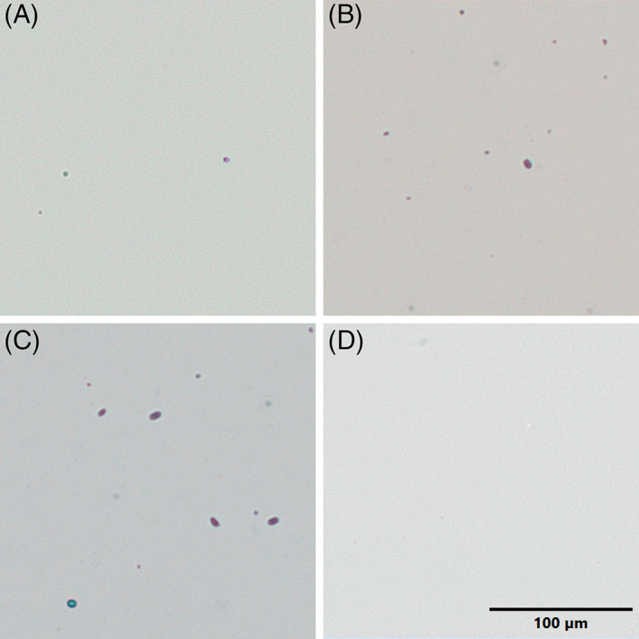
Cellular material extracted using the ZEG. Following ZEG extraction of Xenopus tropicalis embryos, selected samples across a range of DNA concentrations were mixed with Trypan Blue (1:1) and imaged; (A) NF 25 ‐ known DNA concentration 32 ng/μL, (B) NF 25 ‐33 ng/μL, (C) NF 19 ‐ 63 ng/μL, and (D) 0.05X MMR (blank). Few Trypan Blue stained (dead) cells and fragments as well as occasional Trypan‐excluding, presumably live cells, are visible in preparations. Scale bar = 100 μm.