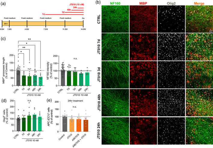
FIGURE 4.

One‐day treatment with JT010 damages myelin in organotypic cortical brain slices, but oligodendrocyte loss only occurs after 7 days exposure. (a) Scheme of organotypic cortical brain slices treatment with JT010 (10 nM) for 7, 3, 2, and 1 days in vitro (DIV). (b) Representative images of 2 weeks organotypic cortical brain slice show immunolabeling for DAPI (blue), NF160 (green), MBP (red), Olig2 (white), and merge in CTRL, JT010 7 days, JT010 3 days, JT010 48 h, JT010 24 h. (c) Quantification of MBP+ process length (left) and NF160+ fluorescence intensity (right; % of control) in CTRL (n = 28, 25), JT010 7 days (n = 11, 12), JT010 3 days (n = 16, 10), JT010 48 h (n = 14, 9), JT010 24 h (n = 12, 9). mean ± SEM, one‐way ANOVA and Bonferroni's multiple comparisons test, *p < .05, **p < .01. (d) Quantification of Olig2+ cells (% of control) in CTRL (n = 23), JT010 7 days (n = 10), JT010 3 days (n = 11), JT010 48 h (n = 8), JT010 24 h (n = 6). (e) Quantification of APC/CC1 + cells (% of control) in CTRL (n = 19), JT010 (n = 17), A967079 (n = 15), and A967079 + JT010 (n = 13). Data are mean ± SEM, unpaired t‐test, **p < .01, ****p < .0001.
