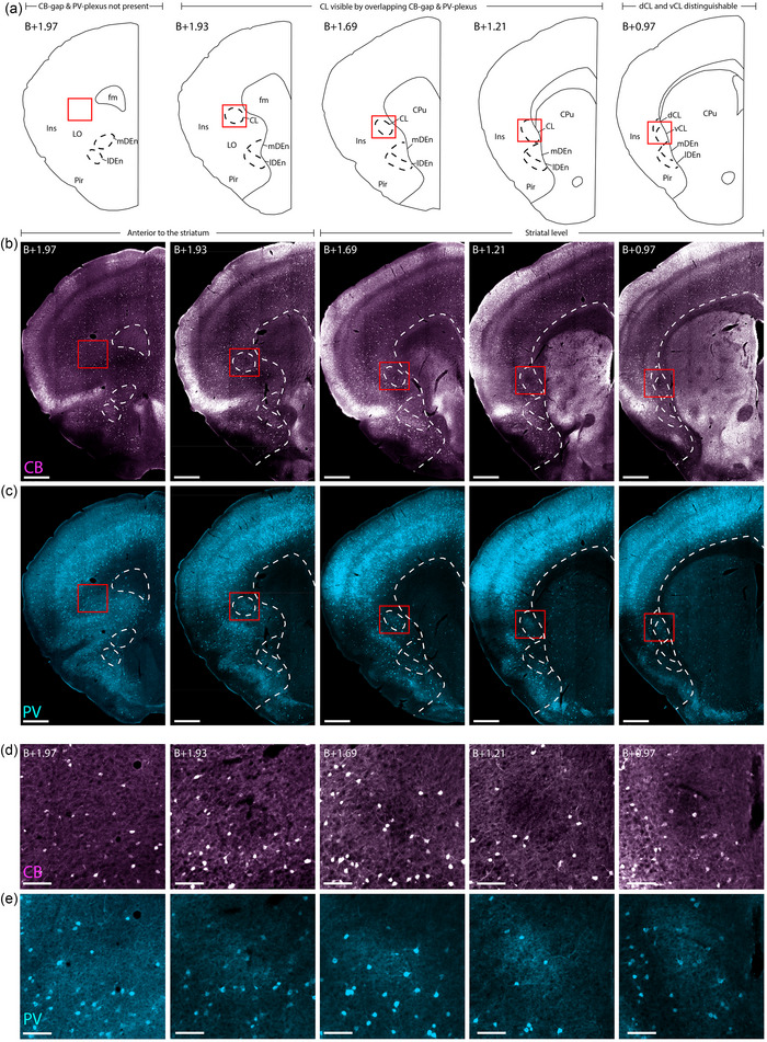
FIGURE 2.

Delineating the rostral‐most parts of the claustrum in female C57 mouse. Immunohistochemical labeling of calbindin (CB) and parvalbumin (PV) in five sections, taken at gradually more rostral levels (right to left, indicated by level rostral to Bregma (B)). (a) Schematic of the histological images shown in (b–e), indicating where the striatum starts and how far rostral the claustrum (CL) can be identified. (b) Location of the CB‐gap, revealing the CL until B+1.93. (c) PV labeling of the same sections as in (b), showing the position of the PV plexus in the CL until B+1.93. (d) Insets from red squares indicated in (b), showing high magnification of the CB‐gap. (e) Insets showing the same areas as in (d) but stained for PV. Note the presence of dense PV neuropil aligned with the CB‐gap. Scale bars measure 500 μm in overview images and 100 μm in insets. All images were obtained with a slide scanner (see Section 2). Abbreviated terms are explained in the list of abbreviations.
