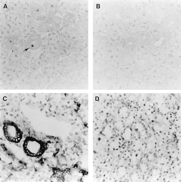
FIG. 10.
Expression of RCMV (early) proteins and rat class II MHC proteins in salivary glands of RCMV- and RCMVΔR33-infected rats. The figure shows micrographs of 4-μm sections of rat salivary glands infected with either RCMV (A and C) or RCMVΔR33 (B and D). Tissue sections were stained with either MAb against viral (E) antigens (MAb RCMV8 [A and B]) or MAb against class II MHC proteins (OX-6 [C and D]). The tissue sections were photographed at a magnification of ×400.

