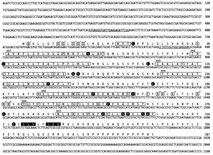
FIG. 2.
Nucleotide sequence of the R33 gene, and predicted amino acid sequence of the pR33 peptide. The open boxes indicate seven putative transmembrane domains (tm1 to tm7) and a putative N-linked glycosylation site (NXT/S). Charged amino acid residues in the N-terminal (extracellular) region and the third intracellular region (between tm5 and tm6) are enclosed in open squares. The charges of these residues are printed at the top right of each square. Black boxes indicate consensus sequences [(S/T)X(K/R)], of which the S/T residue might be phosphorylated by protein kinase C. The amino acid residues that are conserved among all pUL33-like proteins (Fig. 3) are encircled. The residues that are conserved between chemokine receptors (Fig. 3) are enclosed in black circles. The underlined nucleotide sequences indicate sequences identical or complementary to the sequences of oligonucleotides that were used in RT-PCR (Fig. 5B).

