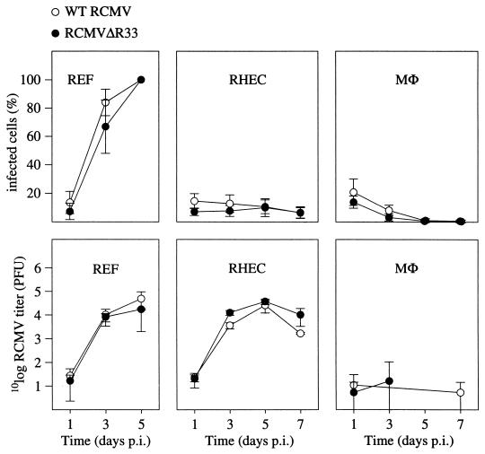
FIG. 8.
The R33 gene is not essential for virus replication in vitro. REF, MHEC, and MΦ were infected with either RCMV or RCMVΔR33, and the replicative potential of these viruses was assessed by immunofluorescence and plaque assay. The upper graphs show the infected-cell/total-cell ratios at various time points p.i. The lower graphs show virus titers that were determined in culture medium up to 7 days p.i. Standard deviations are indicated by vertical bars. REF were monitored up to 5 days p.i., when 100% of the cells showed cytopathic effect. On days 5 and 7 p.i., virus could not be detected in medium samples that were taken from cultures of infected MΦ. Data from these time points are therefore not included in the graph.

