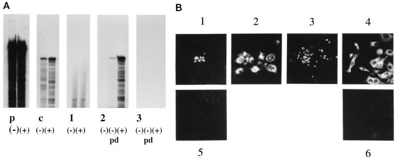
FIG. 2.
Monitoring of BVDV RNA replication. (A) RNase protection assay performed as described in Materials and Methods at 24 h p.t. with MDBK cells transfected with RNA derived from BVDV CP7 cRNA or BVDV CP7Δ3′ cRNA. Lanes: p, input sense (−) and antisense (+) probes (280 nucleotides); c, protected products obtained with sense (−) and antisense (+) probes after RNase protection with BVDV CP7 cDNA; 1, RNase protection carried out with RNA from the nuclear fraction of BVDV CP7 cRNA-transfected MDBK cells (left lane, protection with sense probe; right lane, protection with antisense probe); 2, RNase protection carried out with RNA from the cytoplasmic fraction of BVDV CP7 cRNA-transfected MDBK cells (left lane, protection with sense probe without prior cycle of prehybridization-predigestion (pd); middle lane, protection with sense probe after prior cycle of hybridization-predigestion (pd); right lane, protection with antisense probe); 3, same experiments as in lanes 2 but carried out with cytoplasmic RNA from BVDV CP7Δ3′ cRNA-transfected MDBK cells. The figure is an autoradiogram of a 10% polyacrylamide–7 M urea gel. (B) IF analysis at 24 h p.t. of MDBK cells transfected with either BVDV CP7 cRNA or BVDV CP7Δ3′ cRNA. Anti-NS3 antibody was used. 1, MDBK cells transfected with BVDV CP7 cRNA (magnification, ×100); 2, same as 1 but at a magnification of ×400; 3, different image of MDBK cells transfected with BVDV CP7 cRNA (magnification, ×100); 4, same as 3 but at a magnification of ×400; 5, MDBK cells transfected with BVDV CP7Δ3′ cRNA (magnification, ×100); 6, MDBK cells transfected with BVDV CP7Δ3′ cRNA (magnification, ×400).

