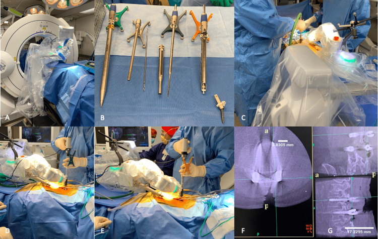
Figure 2. Medtronic Mazor X Stealth Edition intraoperative use during posterior lumbar pedicle screw placement.
(A) Room staging with O-Arm Surgical Imaging System used for intraoperative 2D/3D imaging for integration into the Mazor system and placement of fluoroscopy on the opposing side of the patient for seamless access and transition intraoperatively. (B) From left to right: dilator, midas, drill bit, midas cannula, tap, solera, and MAS driver. The (C) drill, (D) tap, and (E) solera are used sequentially for screw placement. (F) Intraoperative fluoroscopy confirms accurate screw placement on axial imaging. (G) Intraoperative fluoroscopy confirms accurate screw placement on sagittal imaging. In figures F and G, "a" and "p" indicate anterior and posterior orientation, respectively. 'Image Credits: Tania Mamdouhi'.

