
Figure 2. The expression of TGFβ2 in lentiviral injected mouse eyes.
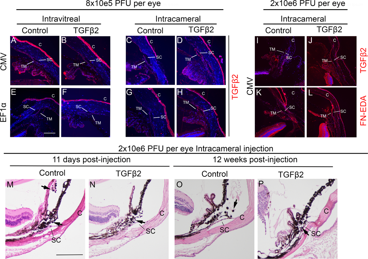
Some mouse eyes were enucleated, fixed, embedded in paraffin, and immunostained for TGFβ2 and/or fibronectin isoform EDA (FN-EDA) (shown in red) and with DAPI (blue). A-H: the eyes that received low viral doses. I-L: the eyes that received high viral doses. Representative images are shown. Blue: DAPI, Red: TGFβ2. Control: eyes injected with either pLVX-CMV-AcGFP or pLVX-EF1α-mCherry lentiviruses. TGFβ2: eyes injected with either pLVX-CMV-ΔhTGFβ2C226S/C228S or pLVX-EF1α-ΔhTGFβ2C226S/C228S lentiviruses. C: cornea, SC: Schlemm’s canal, TM: trabecular meshwork. The white scale bar: 100μm. Some mouse eyes receiving intracameral injection of 2 × 10⁶ pLVX-CMV-AcGFP (M and O) or pLVX-CMV-ΔhTGFβ2C226S/C228S (N and P) were stained with hematoxylin and eosin to determine intraocular inflammation at early (M and N) and late (N and P) time points. SC: Schlemm’s canal, C: cornea, black arrows: inflammatory cells. The black scale bar: 200μm.