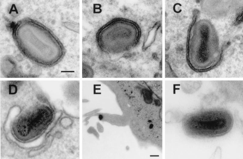
FIG. 9.
Electron microscopy of cells infected with the indicated viruses. Note the formation of intracellular virus particles that have been, or are being, wrapped by intracellular membranes in cells infected with WR WT, vSCR1, vSCR1-2, and vSCR1-3 (A to D). (E) Enveloped particle leaving a B5R/TK-infected cell on an actin tail. (F) EEV particle produced from a vΔB5R-infected cell. Bars: (A to D and F) 100 nm; (E) 500 nm.

