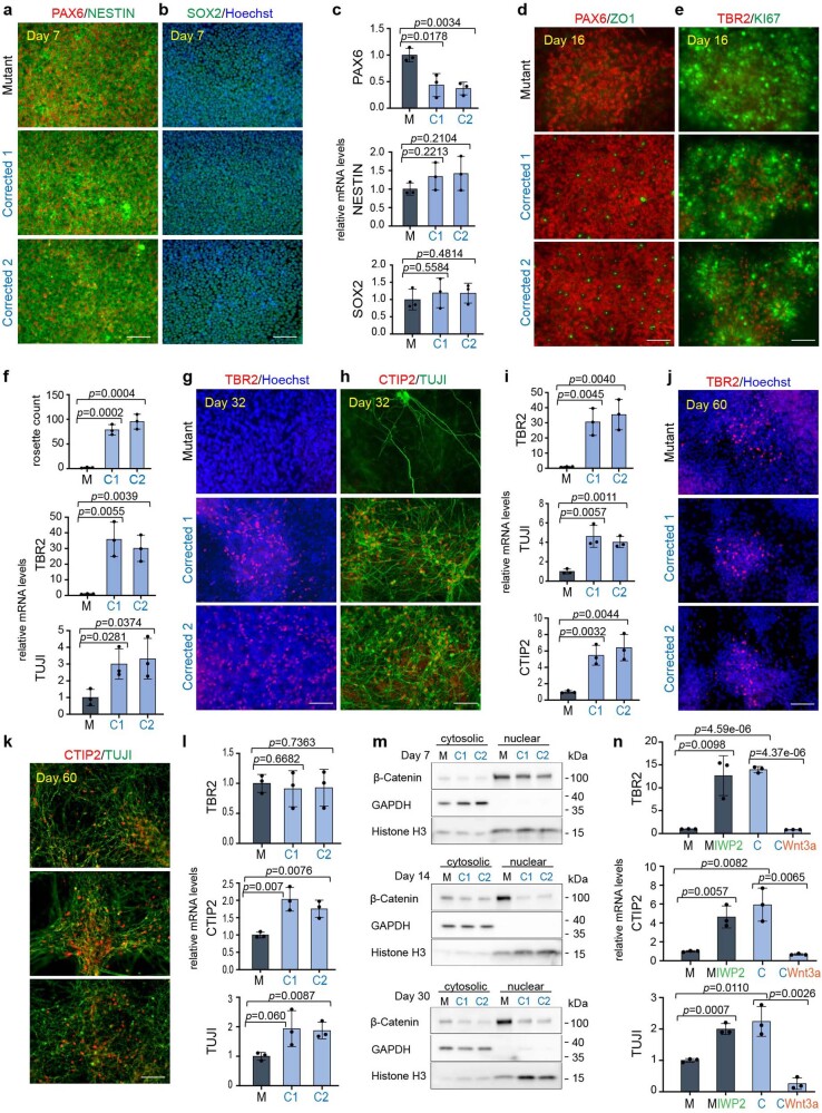
Extended Data Fig. 4. KDM5C mutation leads to a delay in neuronal differentiation and can be rescued and induced by canonical Wnt signaling manipulation in the second brother.
a, Immunofluorescence for PAX6, NESTIN and (b) SOX2 at day 7 of neuronal differentiation in Mutant and two Corrected lines of brother 2. Cells were counterstained with Hoechst. 3 independent experiments were performed with similar results. c, qPCR analysis for PAX6, NESTIN and SOX2 mRNAs at day 7 in the second brother. Data are represented as mean ± SD of 3 independent experiments. The p-values by two-tailed unpaired Student’s t-test are indicated. P < 0.05 was considered statistically significant. d, Immunofluorescence for PAX6, ZO1 and (e) TBR2 and KI67 at day 16 of neuronal differentiation in Mutant and two Corrected lines of brother 2. 3 independent experiments were performed with similar results. f, Rosette count in Mutant (M) and Corrected lines (C1 and C2) (top) and qPCR analysis for TBR2 and TUJI mRNAs at day 16 in the second brother. Data are represented as mean ± SD of 3 independent experiments. The p-values by two-tailed unpaired Student’s t-test are indicated. P < 0.05 was considered statistically significant. g, Immunofluorescence for TBR2 and (h) CTIP2 and TUJI at day 32 of neuronal differentiation in Mutant and two Corrected lines of brother 2. Cells were counterstained with Hoechst. 3 independent experiments were performed with similar results. i, qPCR analysis for TBR2, TUJI and CTIP2 mRNAs at day 32 in the second brother. Data are represented as mean ± SD of 3 independent experiments. The p-values by two-tailed unpaired Student’s t-test are indicated. P < 0.05 was considered statistically significant. j, Immunofluorescence for TBR2 and (k) CTIP2 and TUJI at day 60 of neuronal differentiation in Mutant and two Corrected lines of brother 2. Cells were counterstained with Hoechst. 3 independent experiments were performed with similar results. l, qPCR analysis for TBR2, TUJI and CTIP2 mRNAs at day 60 in the second brother. Data are represented as mean ± SD of 3 independent experiments. The p-values by two-tailed unpaired Student’s t-test are indicated. P < 0.05 was considered statistically significant. m, Western blotting of nuclear and cytoplasmatic fractions in Mutant and Corrected lines of brother 2 at day 7, 14 and 30 of neuronal differentiation. GAPDH and Histone H3 were used to mark the cytosolic and nuclear fraction respectively. β-Catenin expression in the cytoplasm and nucleus is indicated. At day 14, 3 independent experiments, and at day 7 and day 30, 2 independent experiments were performed. GAPDH, Histone H3 and β-Catenin were run on the same gel. For gel source data see Supplementary Fig. 1. n, q-PCR analysis of TBR2, CTIP2 and TUJI mRNAs at day 30 of neuronal differentiation after treatment regime according to Fig. 3e with the Wnt inhibitor IWP2 in Mutant cells and Wnt induction with recombinant Wnt3a in Corrected cells of brother 2. Data are represented as mean ± SD of 3 independent experiments. The p-values by two-tailed unpaired Student’s t-test are indicated. P < 0.05 was considered statistically significant. Abbreviations: M=Mutant; C1 and C2=Corrected 1 and Corrected 2.

