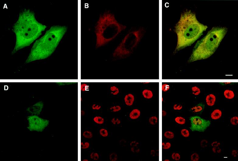
FIG. 7.
Localization of UL32 in cells transfected with pAPVEEUL32. Cells shown in panels A to C were transfected with pAPVUL32 and pVP16 as described under Materials and Methods. Cells shown in panels D to F were transfected with pAPVEEUL32 and superinfected with hr64. In panels A and B, green represents staining with the EE monoclonal antibody and red represents staining with the UL32 polyclonal antibody, respectively. In panel D green represents staining with the EE monoclonal antibody, and in panel E red represents staining for ICP8. Panels C and F show the merged images of the staining patterns. Bars = 10 μm.

