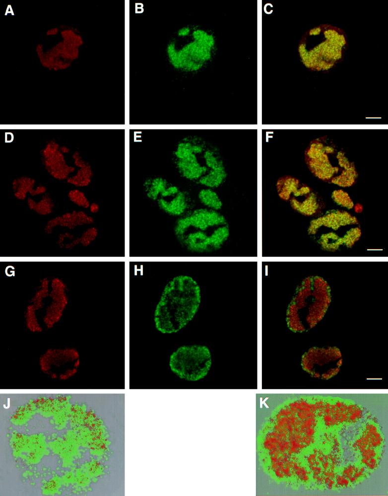
FIG. 8.
Replication compartments and capsid localization. Vero cells were infected with KOS (A to C and J), hr74 (D to F), or hr64 (G to I and K) for 6 h and stained with anti-ICP8 polyclonal antibodies (A, D, and G) and 5C monoclonal antibodies (B, E, and H). Panels C, F, and I represent the merged images of staining patterns with ICP8 and 5C antibodies. Panels J and K represent three-dimensional reconstructions from Z series images of KOS- and hr64-infected Vero cells obtained with the Voxel View program as described under Materials and Methods. In a control experiment, mock-infected cells were treated with primary and secondary antibodies, and infected cells were treated with the secondary antibodies alone; in neither case was any cross-reactivity for anti-ICP8 or monoclonal antibody 5C observed (data not shown). Bars = 10 μm.

