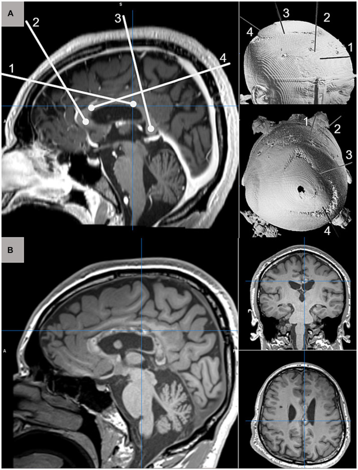
Figure 1.
(A) Schematic depiction of four MRgLITT trajectories for complete CC ablation targeting the genu (1), posterior body (2), anterior body (4), and splenium (3) displayed on sagittal T1-weighted pre-operative MRI (left) and 3D CT reconstruction from anterolateral and superior perspectives. (B) Postoperative T1-weighted MRI acquired 3-months after complete CC ablation demonstrating normal postoperative changes along the extent of targeted callosal white matter.

