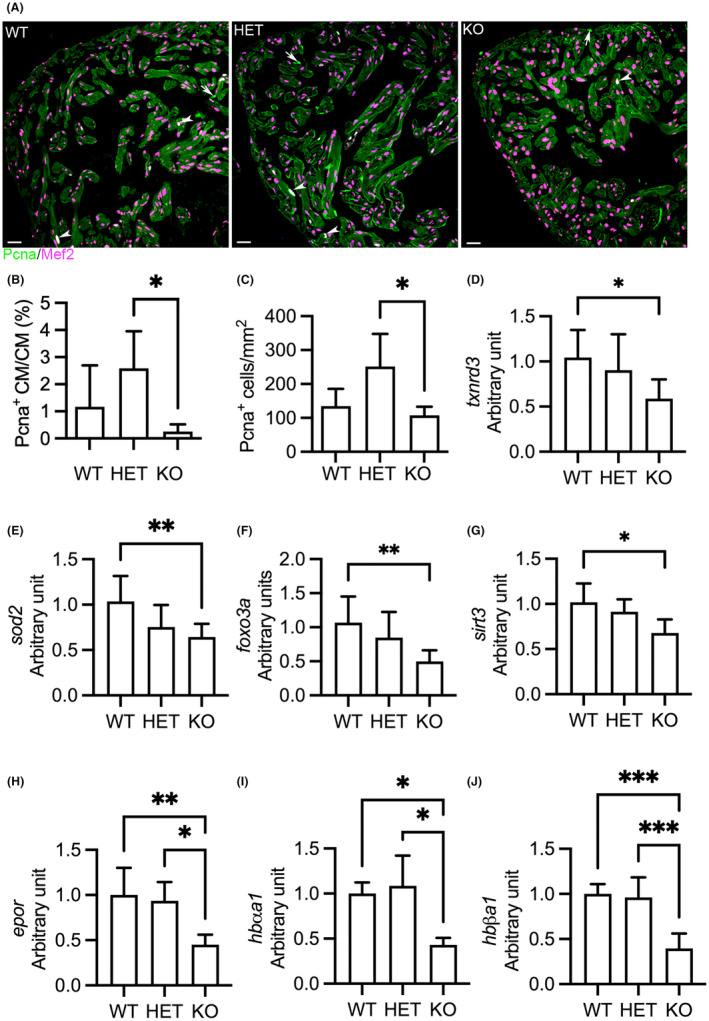
FIGURE 4.

Deletion of hmox1a restrains cardiomyocyte proliferation and downregulates antioxidative genes and haemoglobin genes in adult zebrafish. (A) Representative immunohistochemical staining of Pcna (in green) and Mef2 (in meganne) in ventricular sections. Arrow indicates Pcna+ non‐cardiomyocytes. Arrowhead indicates Pcna+ cardiomyocytes. Pcna, proliferating cell nuclear antigen; Mef2, myocyte‐specific enhancer factor 2. (B) Quantification of Pcna+ cardiomyocytes relative to total cardiomyocytes. (C) Quantification of Pcna+ cardiac cells per mm2 ventricular area. Three hearts from each genotypic group were selected. Two sections from each heart were stained. The number of stain‐positive signals for Pcna and Mef2 from ventricular areas was quantified with ImageJ. (D–G) RT‐qPCR analyses indicating downregulation of antioxidative genes, txnrd3 (D) and sod2 (E), and the transcription regulators of antioxidative and antihypertrophic signalling, foxo3a (F) and sirt3 (G), in KO(52del) hearts. txnrd3, thioredoxin reductase 3; sod2, superoxide dismutase 2; foxo3a, forkhead box O3a; sirt3, sirtuin 3. The graphs represent the quantification of two individual analyses of RNA extracts from pooled samples of two‐three hearts. Each analysis includes three replicates. WT n = 19; HET(52del) n = 19; KO(52del) n = 19. (H–J) RT‐qPCR analyses showing downregulation of erythropoietin receptor epor (H), and adult haemoglobin genes hbαa1 (I) and hbβa1 (J) in kidney lacking hmox1a. epor, erythropoietin receptor; hbαa1, haemoglobin alpha adult‐1; hbβa1, haemoglobin beta adult‐1. The graphs represent the quantification of two individual analyses of RNA extracts from pooled samples of two kidneys. Each analysis includes three replicates. WT n = 10; HET(52del) n = 10; KO(52del) n = 10. Data are presented as mean ± SD. One‐way ANOVA with Tukey adjustment for multiple comparisons. *p < 0.05, **p < 0.01, ***p < 0.001. Scale bars: 20 μm.
